
Under the microscope


On the door of Jim Dixon’s office, there’s an image of a yin-yang symbol, two opposites joined together in the shape of a circle. It’s an image we’ve seen hundreds of times but something about this one makes it unique. “It’s two red blood cells,” says Jim. “It’s an image I took from our electron microscope.” It was the first time he’d ever seen two blood cells joined like that so he captured the image to show his colleagues.
Jim is the Assistant Chief Medical Technologist in the Pathology department at the Montreal Children’s Hospital (MCH), a role he’s held for more than six years. Earlier in his career, Jim worked as a marine biologist after graduating from Mount Allison University in New Brunswick but his interests eventually led him to Dawson College where he studied Medical Technology. After Dawson, he started in Pathology at the Montreal Neurological Hospital (MNH), where he worked for 14 years before coming to the Children’s.
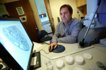
About half of Jim’s time is dedicated to administration duties in the department. The other half involves electron microscopy, or EM, a very specialized area in pathology that the department uses in about 10 percent of the cases they see. There is no formal education for EM—Jim started learning it at the MNH and estimates it took about five years to gain his expertise. Over time, he’s become part of a wider group of people working in this area. “It’s a small field, but we have a strong network across many countries and we often consult with each other,” he says.
An electron microscope differs from other microscopes in several ways. A light microscope, which uses light to help us see an image, can show the size of a strand of hair or a blood cell. Compare that to an electron microscope which can show the size of a virus particle, a DNA sample, or an atom. “The electron microscope at the MCH lets us see images up to 50,000 times larger than they are,” says Jim. “We can actually look at one cell at a time.” Jim estimates that half his time using the electron microscope is devoted to preparing the samples, part of which involves applying heavy metals to give them contrast. “Inside the microscope, there’s no air, humidity, or dust,” says Jim, “only electrons.”
The Pathology department provides services to most departments at the MCH to help diagnose a number of different diseases and conditions. “The majority of cases in pathology are pretty routine,” says Jim, “and can be diagnosed using regular microscopes.” The remaining cases depend on results from the electron microscope. “EM is actually too sophisticated for most cases, but it’s there when we need it.” In one recent case, the electron microscope enabled pathologists to zero in on a particular condition, one that helped rule out the initial diagnosis. “It’s exciting when we can do that,” says Jim. “The results of what we find can sometimes make a major difference in diagnosing a patient’s condition and helping achieve the best possible outcome.”
The MCH Foundation provided funding to purchase the hospital’s electron microscope, which is the only one at the McGill University Health Centre (MUHC). The MCH is the referral centre for electron microscopy at the MUHC and also does work for the Lakeshore General Hospital and St. Mary’s Hospital Center.