A day in the life of three medical technologists in the MUHC Central Lab


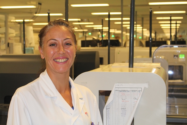
There’s a good chance you’ve never met medical technologists Jessica Driscoll, Annie Malenfant and Rabii El Ouarari. “We definitely work behind the scenes,” says Jessica. The Research Institute of the McGill University Health Centre houses the MUHC’s Central Lab, named for the Marcelle and Jean Coutu Foundation, where the trio work alongside each other to identify, confirm and categorize different types of leukemia in pediatric and adult patients.
Step 1: Reviewing abnormal test results
Childhood leukemia is the most common form of cancer in children and adolescents. It affects a child’s white blood cells and can be detected through a series of tests and careful analysis. Jessica Driscoll’s work involves reviewing abnormal blood results obtained during a complete blood count (CBC) to help identify potential leukemia cases.
A CBC is a blood test used that evaluates a person’s overall health and it can detect a wide range of disorders and diseases, including leukemia. Jessica reviews the patient’s red and white blood cell counts, as well as their platelets and hemoglobins. “I look at the scattergram first, which is a graph that differentiates each white blood cell by cell type and size, and then I confirm my suspicion with the numeric results,” she explains.
Jessica and her colleagues review 150 pediatric CBC results a day. “Thankfully, most of these results end up being non-leukemic diseases or disorders,” she says. Besides helping physicians make a diagnosis, she also reviews blood tests of oncology inpatients and outpatients currently undergoing treatments such as chemotherapy, and patients in the Neonatal Intensive Care Unit and Pediatric Intensive Care Unit who are battling various diseases and disorders.
When searching for signs of leukemia, some of the red flags are very high or very low white blood cell counts, as well as low hemoglobin and platelet levels. “The moment I see a red flag, I immediately get on the phone with the physician who requested the CBC,” she says. “It’s a hard phone call to make, because I know I’m about to completely change someone’s life.”
After informing the physician of her findings, she quickly heads over to the morphology room with a microscope slide in hand. “This is where Annie Malenfant comes into play,” she says. “She takes a look at the smear and if she sees any suspicious cells, she calls the hematologist, who then comes down to the lab to take a closer look and ask for further testing. And of course, the faster we get this done, the better.”
Step 2: Analyzing a patient’s cells
Annie Malenfant spends her days looking at cells through a microscope. She specializes in cellular morphology, which involves identifying the shape, structure, form and size of cells. “I can look at a cell and tell whether or not it’s a cancer cell,” she says. “When reviewing someone’s blood counts, it’s difficult to differentiate between an infection and leukemia, because in both instances, the patient’s white blood cell count is abnormal. But I can tell the difference by looking at the cells.”
When a patient is battling an infection, the body produces a type of infection-fighting white blood cell, called a lymphocyte; and when a patient is battling leukemia, the bone marrow begins to rapidly produce abnormal white blood cells. “Because these white blood cells are produced so rapidly, they tend to look very immature,” she explains. “That’s when I know it could be leukemia.”
Immature cells, also known as blast cells, tend to have a smoother-looking nucleus and their surface is more textured. They also have a different colour. “Cells battling an infection tend to have a darker blue cytoplasm, and they’re reactive. You can actually see them reacting to the environment around them,” says Annie.
On any given day, Annie will analyze 40 to 60 slides, which include blood specimens, bone marrow, as well as cerebral and body fluids. She is also sometimes called onto the unit to help retrieve cells during a bone marrow aspiration. “To get the best quality result, I need to be present in order to make a slide immediately at the child’s bedside,” she says. “At the end of the day, there is always a patient behind each cell, each slide, so we do whatever we can, as quickly as we can, to get a diagnosis.”
Diagnosing leukemia in young children, especially newborns, is especially challenging. “Babies are the most difficult to diagnose, because all of their cells look immature and young. It’s hard to distinguish between a blast cell and a normal, newborn cell. This is why Rabii’s work is so important. He not only helps narrow down the exact type of leukemia, but he can confirm the cases which are difficult to detect with a microscope.”
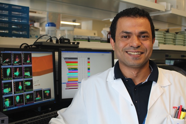
Step 3: Narrowing down the diagnosis
Rabii El Ouarari, a former teacher from Morocco, always loved the idea of working in a laboratory and decided to make his dream a reality after immigrating to Quebec. After completing his degree in medical technology, Rabii started working at the MUHC, and quickly discovered he had an interest and a particular skill for working in hematology.
Rabii specializes in flow cytometry, a laser-based technology used for cell analysis. It allows Rabii to rapidly and accurately profile over 100,000 cells at a time. “I can collect data quickly from thousands of cells, because flow cytometry allows me to view many antibodies at the same time,” he says. “This process can lead to the diagnosis, prognosis and classification of various forms of leukemia.”
In fact, Rabii can detect more than 20 different types of leukemia, which allows physicians to treat their patients with specific therapies targeted to their type of cancer. “There are many different types of leukemia and they all have different characteristics,” he says. “I’m able to distinguish between lymphoid and myeloid leukemias by looking at their distinct cellular features, including their antigens [proteins] and antibodies.”
He is able to make this distinction thanks to a technique called immunophenotyping. This test allows Rabii to identify specific antigens found only in cancerous cells. “The antigens are stained with a fluorescent marker and this marker only binds to specific antigens,” he explains. “This allows me to see the leukemia cells, because they are a different colour from normal cells.”
In 2016, the MUHC became part of EuroFlow, a consortium composed of research centres with expertise in flow cytometry and immunophenotying. “We are now leaders in Canada and are performing at the highest standard,” says Rabii. “We can diagnose different subtypes of leukemia even more precisely than before. It has completely changed our lives and the lives of our patients.”